Diagnosing Pain
Diagnosing Pain
How Doctors Diagnose Pain
If you’re in pain, your doctor has a variety of options for determining what’s causing it. They’ll inquire about your symptoms as well as your medical history, which will include any illnesses, injuries, or surgeries.
In addition to an examination, your doctor may order blood tests or X-rays. The following tests can help you locate the source of your pain:
Computed tomography scan: A CT scan uses X-rays and computers to create an image of a cross-section of the body. You lie on a table as still as possible during the test. It will pass through a piece of big scanning equipment in the shape of a doughnut. Before your scan, your doctor may inject a solution into a vein. It can help you view what’s happening inside more clearly. CT scans typically take 15 to 60 minutes.
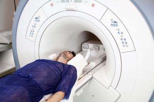
Magnetic resonance imaging: MRI can provide your doctor with clear images without the use of X-rays. This test creates graphics using a huge magnet, radio waves, and a computer. Depending on the number of images taken, an MRI might take anywhere from 15 minutes to over an hour. An injection of contrast material is required for certain MRIs in order to get sharper images. Some people, such as those with pacemakers, should avoid having an MRI since it involves magnets.
Nerve blocks: These tests can help treat and diagnose your pain. A numbing agent (anaesthetic) is injected into nerve locations by your doctor. They might perform an imaging test to figure out where the needle should go. Your reaction to the nerve block could reveal what’s causing your discomfort and where it’s coming from.
Discogram: This exam (also known as Discography) is for individuals who are thinking about having surgery for their back pain. Doctors also utilise it to conduct tests prior to making a treatment decision. A dye is injected into the disc that is suspected to be causing the pain during this test. On X-rays, the dye highlights damaged areas.
Myelogram: This test is also for back pain. A dye is injected into your spinal canal during a myelogram. The test aids in the detection of nerve compression due to herniated discs or fractures.
Electromyogram: Doctors can use an EMG to check muscular activation. Fine needles are inserted into your muscles by your doctor to test their response to electrical signals.
Bone scans: These aid in the diagnosis and tracking of infections, fractures, and other bone problems. A small amount of radioactive material is injected into your bloodstream by a doctor. The debris will collect in the bones, especially in abnormal places. A computer can then identify those exact regions.
Ultrasound scanning: This procedure, which is also known as ultrasound imaging or sonography, employs high-frequency sound waves to create images of the inside of the body. The echoes of sound waves are recorded and shown in real-time.


